For more than six decades photographer Jan Hinsch has been creating beautiful images using his camera and his microscope. He has had numerous exhibitions of his work and for forty-seven years was on the staff of E. Leitz in the company’s microscopy division. His studio in northern New Jersey reflects an enduring love of photography as well as an embrace of science and precision optics. Above his worktable and his Leitz microscopes hangs a favorite image by Henri Cartier-Bresson. In creating his images Hinsch avoids computer manipulation and uses only procedures that are standard in microscopy, such as staining and the use of polarized light—techniques which he uses masterfully. In his continuing exploration of the microscopic world, he is currently experimenting with the iridescent colors and patterns of Wild Turkey feathers he has found on walks near his home. Below the thumbnails Jan Hinsch provides some interesting details on the images.
click image to enlarge — use arrows to advance — use escape key or a vertical swipe to return here
Legend: Eight Photomicrographs
Calcite, Augite, Feldspar — A thin slice of rock, about 30 um thick, consisting of the minerals calcite, augite, feldspar. Micro technique: Low power. Transmitted, polarized light.
Sulfur — Sulfur, heated to the melting point between two pieces of glass, photographed after re-crystallization. Micro technique: Low power. Transmitted, polarized light.
Mixed Diatoms — Diatoms are a group of marine and freshwater algae that form silica cell wall, called frustules, which are depicted here. Micro technique for Mixed Diatoms: Low power transmitted light brightfield. Several pictures were taken at different focus positions so that all appear simultaneously in focus after combining them by software (Combine Z by Alan Hadley.)
Diatoms — Diatoms are a group of marine and freshwater algae that form silica cell walls, called frustules, which are depicted here. The blue appearance of some fragments of frustules are due to their repeating structure elements, which are spaced at distances of the order of the wavelength of light. Micro technique for Diatoms: Medium power transmitted light, Differential Interference Contrast (DIC). This enhances the contrast and the saturation of the blue structures.
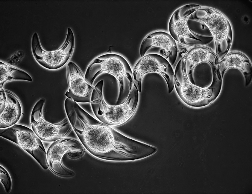
Pyrocystis lunula — These one-celled planktonic plants were collected in the Caribbean. When stimulated they tend to give off flashes of green light, hence the name “moon-shaped fire bag”. Under laboratory conditions one can dramatically demonstrate the presence of chlorophyll in the center of each individual by fluorescence microscopy. Even without the fireworks this is a plant of striking geometry and beauty. Micro technique: Low power, transmitted light phase contrast to enhance the contrast of the organelles.
Sitka — Thin section of the wood of a sitka tree. Micro technique: Low power transmitted light. After cutting, the section is stained with a red dye and observed using polarized light to enhance the contrast.
Trichodina fultoni — A fish parasite. Micro technique: Medium power, transmitted light DIC.
Diamond, Trigons — Natural surface of a raw diamond. Micro technique: Low power, reflected light DIC, which accentuates the trigons and is also the reason for the colors on the left where the surface drops off rather steeply.


